Thursday, October 29, 2026

Human African trypanosomiasis

Challenges to Elimination of gambiense-HAT and Comparison with rhodesiense-HAT

Human African trypanosomiasis (HAT), also known as sleeping sickness, is caused by two related parasites of Trypanosoma brucei. The parasites are transmitted by tsetse flies and has historically posed a major public health burden in sub-Saharan Africa. The chronic form, T. b. gambiense (gambiense-HAT), accounts for the vast majority of cases is predominant in the Western and Central Africa while T. b. rhodesiense is more frequent in Eastern and Southern Africa. Although these fatal infections (Untreated cases) are in the decline trend, the elimination of gambiense HAT is still challenging.

Democratic Republic of the Congo (DRC) has set a goal to eliminate the transmission of gambiense HAT by 2030. Since the cases are declined in the recent years, several biological, epidemiological, and operational challenges complicate the path toward elimination. These challenges relate to the existence of human and animal reservoirs, limitations in surveillance, and constraints in diagnosis, treatment, and integrated vector control. One health approach should be implemented to achieve the goal to eliminate by the target year. This integrated approach includes the control of human and animal reservoirs, extensive surveillance strategy, and control strategies on diagnosis, treatment and vector.


1. Human and Animal Reservoirs

Human is the primary reservoir that plays a key role in transmission of gambiense-HAT disease making a major concern for control strategy. In addition, studies suggest that asymptomatic human carriers may harbor parasites for extended periods without detectable parasitemia using standard diagnostics. These individuals can continue transmission of this infection silently, acting as “cryptic reservoirs” (Buscher et al. 2018). These infections even challenge for elimination because they escape the routine screening programs leading undiagnosed.

Likewise, gambiense HAT has animal reservoirs as well. Animal reservoirs—including pigs, dogs, and other domestic or wild animals—have been found to carry T. b. gambiense in some endemic areas. Although their epidemiological significance remains unclear, experimental and field data indicate that animals could contribute to transmission cycles, particularly in regions where human cases are declining (Mehlitz et al., 2019). If animals maintain the parasite independently of humans, elimination strategies focused solely on human case detection may be insufficient. The unpredictability of transmission cycles, including human and potential animal reservoirs, underscores the warning to “expect the unexpected” (Mehlitz et al., 2019). These hidden reservoirs represent a major obstacle to achieving transmission chain blockage.


2. Surveillance Strategies

The surveillance of entire communities has been highly successful in reducing disease burden during outbreak and higher number of cases using traditional active screening campaigns. However, a number cases are dropping, an alternative, sustainable and effective control strategy is utmost which is logistically challenging. As a consequence, health care center based screening is an alternative cost effective option. This method shifts toward passive surveillance, is necessary but presents difficulties. Lacking proper training for healthcare workers, resources and facilities at health care centers may miss the recognition of parasites and diagnosis gambiense HAT, especially when encounter with patients showing nonspecific early symptoms. Moreover, reduced awareness as cases decline can lead to underreporting and delayed diagnosis.

New approaches include:

  1. Targeted surveillance, focusing on high-risk populations and historical foci
  2. Serological screening tools (e.g., rapid diagnostic tests) to identify suspected cases
  3. Molecular diagnostics, which may detect low-level infections even in early stage of infection but remain expensive and technically demanding

Additionally, xenomonitoring (presence of parasites in tsetse flies) may be used as supplementary screening strategy to detect ongoing transmission (Rotureau et al., 2013). However, implementing such methods at scale remains challenging. A major issue is maintaining sensitive surveillance systems in low-prevalence settings.


3. Control Strategies

Diagnosis

The diagnosis of gambiense-HAT is based on a two-step process: serological screening followed by parasitological confirmation. Although the method is effective, it has few limitations including low sensitivity while detecting low parasitemia infections. In addition, skilled manpower and well facilitated laboratory infrastructure are required. The most important challenge is identifying latent infections or infections with non-specific symptoms. Recent developments, such as oral treatments that may not require parasitological confirmation, could simplify diagnostic pathways, but their integration into elimination programs requires careful evaluation, particularly in the context of evolving control strategies and uncertainties around transmission dynamics and reservoirs (Mehlitz et al., 2019; Buscher et al.).


Treatment

The introduction of safer and simpler drugs improves the treatment significantly. Oral therapies of antiparasitic drug; fexinidazole allow the effective treatment of a number of outpatients reducing logistical burdens. However, ensuring accessibility to remote areas, managing treatment adherence and avoiding drug resistant remain still challenging. Furthermore, treating asymptomatic carriers—if identified—raises ethical and operational questions.


Integrated Vector Control

The tsetse fly is the sole vector of HAT, making vector control a critical component of elimination strategies. A multiple approach is necessary to control the vectors that not only includes tsetse fly biting but also reduce their population. The use of insecticide-treated targets and traps, aerial spraying and sterile insect technique (SIT) are some of those methods used for vector control. Recent innovations such as “tiny targets” have proven highly cost-effective in reducing tsetse populations. However, sustaining vector control over large and often inaccessible areas is difficult. Ecological factors, such as tsetse habitat diversity and reinvasion from untreated areas, complicate efforts (Van den Bossche et al., 2011). The complicity of transmission dynamics of gambiense HAT also stays major obstacle on vector control.

The transmission efficiency of the vector depends on vector competence that is influenced by complex biological interactions within the fly, including different parasite development stages (procyclic forms, metacyclic forms) (Rotureau et al., 2013).


Would the same strategy work for rhodesiense-HAT?

The above elimination strategy used for gambiense-HAT may not be directly applicable to T. b. rhodesiense (rhodesiense-HAT), which differs fundamentally in its epidemiology. Rhodesiense-HAT is an acute zoonotic disease, primarily infecting Eastern and Southern Africa that accounts for only 8% of reported cases. The number of cases are also much less than gambiense-HAT. In addition, wildlife and livestock serving as the primary reservoirs for rhodesiense-HAT, rather than humans gambiense-HAT. Transmission occurs at the interface between wildlife, livestock, and human populations (Auty et al., 2016). Consequently, humans are incidental hosts, not the main reservoir. Only eliminating human cases does not interrupt transmission, rather animal reservoirs sustain the parasite independently and maintain within the animal population. Hence, a slight different approach should be implemented to eliminate rhodesiense-HAT transmission.

Control strategies must therefore focus on integrated approaches, focusing on treatment and proper management of livestock as cattle are major reservoirs. Vector control in livestock and wildlife areas are essential although vector control in wildlife is the most challenging one. Moreover, the physical barrier for human-animal interfaces should be clearly defined to avoid close interactions.

The concept of a Progressive Control Pathway for African Animal Trypanosomosis (AAT) highlights the need for stepwise, multisectoral strategies targeting animal reservoirs (Diall et al., 2017).

Unlike gambiense-HAT, where elimination of transmission is theoretically feasible due to the predominance of human reservoirs, rhodesiense-HAT control is more about reducing incidence rather than complete elimination. The ecological complexity and involvement of wildlife make eradication extremely difficult.


Concluding remark

The decline of gambiense-HAT in the DRC is a landmark public health achievement, yet reaching the 2030 goal of zero transmission remains a demanding effort. There is a complex set of hurdles to face: "silent" or cryptic human infections that go undetected, the looming possibility of animal reservoirs, and the logistical difficulty of maintaining robust surveillance and treatment in high-risk areas. Overcoming these barriers will require more than just persistence; it demands flexible strategies and a new generation of diagnostic and vector control tools. A one health approach should be adopted that treats human, animal, and environmental health as one interconnected system where all stakeholders work together to achieve the goal.

The situation with rhodesiense-HAT, however, is a different story entirely. Because this form of the disease is zoonotic—meaning it cycles through animals—human-focused medical care alone won't be enough. To make a real impact, only treating infected human population may not work, rather control of infections in wild and domestic animals within their natural habitats may be the mainstay. Even then, given the current tools at our disposal, complete elimination remains a distant and difficult target.


Adopted from CDC


Saturday, September 12, 2026

High burden of blaKPC−2–positive carbapenemresistant Klebsiella pneumoniae among inpatients in a Tertiary Care Hospital in Kathmandu


High Burden of blaKPC-2–Positive Carbapenem-Resistant Klebsiella pneumoniae among Inpatients in a Tertiary Care Hospital in Kathmandu

Shashi Gurung1, Milan Kumar Upreti1, Agrani Paudel2, Khadga Bikram Angbuhang1, Sanjit Shrestha3, Upendra Thapa Shrestha2 *, Megha Raj Banjara2 *

1 GoldenGate International College, Department of Microbiology, Battisputali, Kathmandu, Nepal

2 Central Department of Microbiology, Tribhuvan University, Kirtipur, Kathmandu, Nepal

3 Department of Pathology, Kirtipur Hospital, Kathmandu, Nepal


*Correspondance: Dr. Megha Raj Banjara, Professor; Dr. Upendra Thapa Shrestha, Assistant Professor, Central Department of Microbiology, Tribhuvan University, Kirtipur, Kathmandu. Email:  megha.banjara@cdmi.tu.edu.np/upendra.thapashrestha@cdmi.tu.edu.np/

Abstract

Objective Klebsiella pneumoniae, a major nosocomial pathogen, is increasingly associated with multidrug resistance and carbapenem production, posing a significant therapeutic challenge on a global scale. The study aimed to determine the distribution, antimicrobial susceptibility pattern, and molecular identification of blaKPC-2 and blaVIM genes among carbapenem-resistant K. pneumoniae isolated from clinical specimens.

Methods Between January and June 2024, a hospital-based cross-sectional study was carried out at Kirtipur Hospital in Nepal. Standard microbiological protocols were adopted for the processing of 5002 clinical samples. Antimicrobial susceptibility testing was performed using the Kirby–Bauer disk diffusion method following CLSI guidelines. The modified carbapenem inactivation method (mCIM) and the EDTA-modified carbapenem inactivation method (eCIM) were used to confirm carbapenemase production phenotypically, and the polymerase chain reaction (PCR) was used to detect the blaKPC-2 and blaVIM genes at the molecular level.

Results Of the total samples, 18.3% (917) showed bacterial growth, with K. pneumoniae accounting for 7.6% (n=70) of isolates, predominantly from wound/pus (35.7%), urine (32.8%), and blood (17.1%) specimens. High resistance was observed against imipenem (68.6%), ceftazidime (64.3%), and ceftriaxone (62.9%), whereas tigecycline and doxycycline remained the most effective ones. Twelve (30.8%) of the 39 carbapenem-resistant isolates were determined to be carbapenemases producers, all harboring blaKPC-2 (100%) and three (25%) additionally carrying blaVIM.

Conclusion The dominance of KPC-mediated resistance among the hospitalized patients, underscores the need for rigorous infection control, ongoing molecular surveillance, and prudent antimicrobial stewardship to stop the spread of high-risk K. pneumoniae strains in healthcare environments.

Keywords: Klebsiella pneumoniae, Carbapenem-resistant, blaKPC-2, blaVIM


Full Text: Download


Saturday, August 15, 2026

Occurrence of fimH gene in Escherichia coli isolates from urinary tract-infected patients (Next Research)


 Occurrence of fimH gene in Escherichia coli isolates from urinary tract-infected patients

Shekhar Kumar Jhaa, Milan Kumar Upretia, Sanjit Shresthab, Anup Muni Bajracharyac, Anjali Rawald, Upendra Thapa Shresthad *

a GoldenGate International College, Department of Microbiology, Battisputali, Kathmandu, Nepal

b Department of Pathology, Kirtipur Hospital, Kathmandu, Nepal

c Department of Microbiology, Balkumari College, Narayangarh, Chitwan, Nepal

d Central Department of Microbiology, Tribhuvan University, Kirtipur, Kathmandu, Nepal


* Corresponding author: Dr. Upendra Thapa Shrestha, Assistant Professor, Central Department of Microbiology, Tribhuvan University, Kirtipur, Kathmandu, Nepal. Email:  upendra.thapashrestha@cdmi.tu.edu.np / upendrats@gmail.com


ABSTRACT

Background: Urinary tract infections (UTIs) remain a major global public health concern, with Escherichia coli accounting for most community-acquired cases. The pathogenicity of uropathogenic E. coli (UPEC) is closely linked to virulence factors such as Type 1 fimbriae, encoded by the fimH gene, which promotes adhesion to uroepithelial cells. This study investigates the prevalence of the fimH gene in E. coli isolates recovered from UTI patients and examines its association with extended-spectrum β-lactamase (ESBL) production and multidrug resistance (MDR).

Methods: A hospital-based cross-sectional study was conducted from January to July 2022 at the Department of Microbiology, Kirtipur Hospital, Kathmandu, Nepal. A total of 1200 mid-stream urine samples were processed during the study period to isolate and identify the bacterial pathogens, including E. coli. The antimicrobial susceptibility patterns of E. coli isolates were performed by using modified Kirby-Bauer disc diffusion testing following CLSI guidelines. Phenotypic confirmation of ESBL production was performed using the Double Disc Synergy Test. Genomic DNA was extracted using the phenol: chloroform method, and the fimH gene was amplified by conventional PCR using specific primers.

Results: Among 1,200 urine samples analyzed, 330 (27.5%) showed significant bacterial growth, with E. coli representing 293 (88.8%) of isolates. Among the first-line antibiotics tested, E. coli showed the highest resistance to amoxicillin and ceftazidime, while the highest resistance was observed to tigecycline among second-line agents. In contrast, resistance to imipenem, cefoperazone-sulbactam, and piperacillin-tazobactam was limited, with no resistance to polymyxin B and colistin. ESBL production was detected in 96 (32.8%) of E. coli strains. ESBL-producing isolates exhibited significantly higher resistance to ceftazidime, levofloxacin, ofloxacin, nitrofurantoin, and imipenem than non-ESBL producers.  57% of the UPEC were MDR strains. PCR analysis revealed that 70.8% of ESBL-producing isolates and 84.4% of MDR strains carried the fimH gene.

Conclusion:  Escherichia coli was the predominant uropathogen, with a high prevalence of ESBL production and multidrug resistance, while nitrofurantoin and amikacin remained the most effective antimicrobial agents. The fimH gene was highly prevalent among UPEC isolates, particularly in ESBL-producing and MDR strains, providing preliminary molecular epidemiological data on circulating UPEC strains in Nepal.

Keywords: Uropathogenic E. coli, ESBL, Multidrug resistant, fimH


Download: Full Text


Thursday, July 30, 2026

Critical Reflection on the (Re-)Emergence of Rift Valley Fever

Introduction

Before knowing the driving forces for the emergence and re-emergence of Rift Valley fever (RVF) in Africa, we should know about the disease, transmission cycle, and epidemiology. RVF is a mosquito-borne zoonosis caused by a Phlebovirus of the Bunyaviridae family. The virus affects both livestock and humans, with severe implications for public health, food security, and trade. Most importantly, the virus is carried by more than 53 species of mosquitoes in 8 genera within the family Culicidae in regions of epizootics (Linthicum et al., 2016). Since its first description in Kenya in 1931, RVF has exhibited episodic outbreaks across Africa and occasional expansion into the Arabian Peninsula, highlighting its persistent and dynamic epidemiological profile (Nanyingi et al., 2015; Himeidan et al., 2014). Its transmission involves complex interactions between vectors, hosts, and environmental conditions, making it a quintessential One Health challenge. Based on the literature provided in the study materials, the important driving forces were critically examined and assessed its potential for spread beyond Africa. In addition, it also evaluates current control strategies and identifies key unresolved challenges.


The drivers of RVF emergence and re-emergence in Africa are ranked based on the criteria, including 1. Magnitude of impact on outbreak initiation and scale, 2. Strength and consistency of empirical evidence across outbreaks, 3. Geographic and temporal generalizability, and 4. Potential for intervention (modifiability)

1. Climatic and environmental variability (primary driver)

The top-ranked driver for the emergence of RVF from the different literature identifies abnormal rainfall and flooding as the most critical driver of RVF outbreaks. These conditions promote hatching of dormant Aedes mosquito eggs and amplification of vector populations (Linthicum et al., 2016). The massive increased of infected mosquitoes leads to high epizootic potential. In addition, a couple of studies on satellite-based vegetation indices and rainfall anomalies have consistently predicted outbreaks in East Africa (Anyamba et al., 2002). Nanyingi et al. (2015) demonstrate that nearly all major RVF outbreaks correlate with El Niño–Southern Oscillation (ENSO) events. Floodwater Aedes species act as primary vectors, initiating transmission cycles, while Culex species amplify epidemics.

The climatic and environmental variability was considered a primary driver because it was the strongest predictive evidence. It is also directly related to outbreak initiation (Bird and McElroy, 2016).


2. Livestock dynamics and husbandry practices

Another important driver of the emergence of RVF is livestock dynamics. It plays a central role as an amplification host. The high density of infected mosquitoes amplifies virus activity among the livestock within 6-8 weeks. High-density herds, animal movement, and trade networks facilitate rapid viral spread (Balenghien et al., 2013). Consequently, the intensification of livestock production and transboundary trade increase exposure risks to humans. A review of studies from African countries emphasizes socioeconomic practices such as communal grazing, seasonal migration, and informal markets, which enhance virus dissemination (Muga et al., 2015). Additionally, susceptible livestock populations lacking immunity can trigger explosive outbreaks.

Livestock dynamics is considered another important risk factor since it is essential for epidemic amplification and strongly linked to outbreak severity; however, it can be overcome with strict policy and vaccination of livestock.


3. Weak surveillance and health systems

Besides climatic factors and livestock dynamics, the health surveillance system has a direct role in the emergence of RFV. The poor health system is responsible for delayed detection and response that significantly exacerbate RVF outbreaks. Bird and McElroy (2016) highlight gaps in diagnostic capacity, underreporting, and fragmented veterinary-public health coordination as important causes of outbreaks. In addition, limited early warning systems and inadequate integration of animal and human health surveillance delay interventions, allowing outbreaks to expand uncontrolled.

The surveillance health system is critical for outbreak control rather than initiation. The system can be modified and improved; however, it is inconsistently implemented in African countries.


Interconnection between the drivers

These drivers operate synergistically rather than independently. The climatic factor amplifies vector proliferation, which ultimately leads to livestock infections. Consequently, the increase in livestock infections spills over to the human population, causing outbreaks. Likewise, weak surveillance delays detection of climate-driven outbreaks. Lastly, livestock movement spreads infection beyond environmentally suitable zones. For example, heavy rainfall may initiate an outbreak, but livestock trade determines its geographic spread, while poor surveillance allows it to escalate.


Alternative ranking and counterargument

An alternative perspective places socioeconomic and cultural factors as the primary driver (Muga et al., 2015). Human behavior—such as slaughter practices and consumption of raw animal products—directly influences transmission. Sociocultural factors shape transmission intensity but are less predictive of outbreak initiation.


2. Conditions for RVF spread beyond Africa

The conditions for RVF to spread and become established on other continents require three distinct stages, including introduction of virus, local transmission, and establishment.

2.1. Introduction of virus

The introduction of the virus to the human population may be via infected livestock trade (legal or informal) and the movement of infected humans or vectors. The livestock trade is a main cause of the 2000 outbreak in Saudi Arabia and Yemen (Himeidan et al., 2014). The lack of proper regulation and surveillance during trade with endemic regions and weak border security intensifies the introduction of the virus from one region to another or one country to another. International trade plays a crucial role in establishing national animal health policies because it requires countries to maintain and demonstrate high standards of animal health in order to safely import and export animals and animal products.


2.2. Local Transmission

Local transmission is directly associated with a competent mosquito population and suitable climatic conditions as mentioned above. The massive increase in infected mosquito spread virus to livestock, and ultimately spills over to humans. Several mosquito genera are capable of RVF transmission (e.g., Aedes, Culex) and are globally distributed (Elliott & Brennan, 2014). Thus, competent mosquito vectors, suitable climatic conditions, and the presence of susceptible hosts are responsible for local transmission of the disease.


2.3. Sustained circulation (establishment)

Long-term establishment of the infection requires persistent infected mosquito populations, vertical transmission of virus in mosquitoes and continuous availability of susceptible hosts. Bird and McElroy (2016) note that maintenance mechanisms outside Africa remain uncertain, particularly regarding overwintering of the virus.


Are the same drivers relevant outside Africa?

The same drivers may not be entirely relevant outside Africa. Although the climatic factor remains on the top for emergence of RVF, the temperate regions (low temperature range) in many countries may limit vector survival. Likewise, livestock systems differ, often with stronger regulation and biosecurity in other countries playing a role in preventing the spread of the disease. Lastly, stronger surveillance systems are generally reducing outbreak scale, although other factors favor the outbreak. Thus, while climate remains a key driver, trade and biosecurity may become more critical determinants outside Africa.


3. Appraisal of current RVF control strategies

3.1 Diagnostics

The diagnostic approaches of RFV not only detect the disease in the early phase but also help to prevent further spread by breaking the transmission chain. Recent advances such as molecular tools (RT-PCR) enable early and rapid detection within a few hours during the incubation phase, while serological assays with a focus on antibody detection identify past exposure and seroprevalence among the livestock and human population. However, the RT-PCR tool is limited in endemic regions, and serology can’t differentiate between active vs past infection (Bird & McElroy, 2016). Although culture is the gold standard method for diagnosis of viral infection, including RVF, only a few reference laboratories have this facility.


3.2 Surveillance

Integration of satellite data for early warning (Linthicum et al., 2016) and increasing adoption of One Health surveillance systems are key strengths of the surveillance system. However, underreporting and delayed response may hinder control strategies.


3.3 Vaccination

Currently, recommended vaccines (live-attenuated and inactivated vaccines) exist for livestock and humans to control RVF. The new-generation vaccines are under development (Kortekaas, 2014). However, there are a few challenges to using those vaccines. The safety concerns (e.g., teratogenic effects) of new vaccines, their limited availability, and logistical barriers for massive vaccinations are key hurdles in preventive measures of RVF.

3.4 Integrated vector control strategy

Integrated vector control remains key in controlling the spread of infections. Integrated vector control includes the use of insecticides, environmental management to prevent vector breeding, farm sanitation to avoid mosquito bites to livestock, etc. The limitation of vector control is difficulty in sustaining and limited effectiveness during large-scale outbreaks (Balenghien et al., 2013).


3.5 Outbreak preparedness and response

The outbreak preparedness and response team recognizes the importance of One Health approaches to timely control of potential outbreaks and immediate management. It also improves forecasting tools. Sometimes, poor coordination between sectors and insufficient funding and infrastructure may be a barrier to the role of the team in control measures.


4. Conclusion: Unresolved challenges and priorities

Despite advances, several critical challenges remain in controlling RVF. These are;

i. Predictive uncertainty: Although climate-based models are useful, they cannot fully predict outbreak timing or scale.

ii. Vaccine gaps: Safe, affordable, and widely deployable vaccines remain limited. Many vaccines are under development for livestock and humans. These vaccines are in the clinical trial phases. Development costs and licensing costs are still challenges for veterinary vaccines and human vaccines. Only a combined effort from veterinary vaccines to develop and approve human vaccines can reduce costs and licensing procedures.

iii. Poor surveillance system: Persistent gaps in early detection hinder rapid response. In addition, political influences and lack of a single platform to coordinate between stakeholders remain major obstacles in data sharing and policymaking.

iv. Socioeconomic barriers: Although socioeconomic barriers don’t have a direct role in outbreaks and massive transmission, the cultural practices and economic constraints limit intervention uptake. If such conditions are revealed in an endemic region, it may cause further spread of infections and make control measures difficult.


Priority actions

Based on the above information, the following urgent priorities are identified:

  1. We should strengthen integrated One Health surveillance systems that collect data from humans, livestock, and the environment (climate data) for prediction of potential outbreaks.
  2. Secondly, more research should focus on the development of safe and effective livestock vaccination.
  3. Thirdly, the local authorities should focus on an integrated vector control strategy.
  4. Lastly, based on integrated data, we should improve quick data sharing and predictive modeling through interdisciplinary data.  

These actions address both biological and systemic drivers, offering the greatest potential impact.


References

Anyamba A, Linthicum KJ, Mahoney R, Tucker CJ, Kelley PW. 2002. Mapping potential risk of Rift

Valley fever outbreaks in African savannas using vegetation index time series data. Photogramm. Eng. Remote Sens. 68:137–45

Balenghien, T. et al. (2013) ‘Towards a better understanding of Rift Valley fever epidemiology in the south-west of the Indian Ocean’, Veterinary Research, 44(1), pp. 78.

Bird, B.H. and McElroy, A.K. (2016) ‘Rift Valley fever virus: Unanswered questions’, Antiviral Research, 132, pp. 274–280.

Elliott, R.M. and Brennan, B. (2014) ‘Emerging phleboviruses’, Current Opinion in Virology, 5, pp. 50–57.

Himeidan, Y.E. et al. (2014) ‘Recent outbreaks of Rift Valley fever in East Africa and the Middle East’, Frontiers in Public Health, 2, pp. 169.

Kortekaas, J. (2014) ‘One Health approach to Rift Valley fever vaccine development’, Antiviral Research, 106, pp. 24–32.

Linthicum, K.J., Britch, S.C. and Anyamba, A. (2016) ‘Rift Valley Fever: An Emerging Mosquito-Borne Disease’, Annual Review of Entomology, 61, pp. 395–415.

Muga, G.O. et al. (2015) ‘Sociocultural and economic dimensions of Rift Valley fever’, American Journal of Tropical Medicine and Hygiene, 92(4), pp. 730–738.

Nanyingi, M.O. et al. (2015) ‘A systematic review of Rift Valley Fever epidemiology 1931–2014’, 

Saturday, May 23, 2026

Detection of carbapenem resistance and its attributable genes in Acinetobacter baumannii isolated from cardiac patients at a referral cardiac hospital of Kathmandu


Abstract

Objective: Acinetobacter baumannii has emerged as a major nosocomial pathogen due to its remarkable ability to develop resistance to multiple antibiotics including carbapenems. The objective of this study was to assess the carbapenem resistance and detect carbapenem-resistant genes in clinical isolates of A. baumannii.

Methods: A cross-sectional study was conducted in Shahid Gangalal National Heart Centre, Kathmandu during February to September 2024. A total of 42 A. baumannii were isolated from different clinical specimens and identified. Antibiotic susceptibility test was performed by Kirby-Bauer disc diffusion method and carbapenemase production was assessed using modified carbapenem inactivation method and EDTA-carbapenem inactivation method. Carbapenem-resistant genes were detected through polymerase chain reaction. Among 1607 samples tested, 349 were culture positive for bacteria.

Results: The prevalence of A. baumannii was 12% (42/349). Among the 42 A. baumannii isolates, 88.1% were resistant to carbapenems. Metallo-β-lactamase production was observed in 35.7% and multidrug resistance in 83.3% isolates. Resistance rates were highest against cefotaxime, cefepime and carbapenems. The blaOXA-23 gene was detected in 69.1% of the isolates, blaNDM-1 in 66.7%, and blaVIM in 14.3%, but none of the isolates harbored the blaIMP. Co-occurrence of blaOXA-23 and blaNDM-1 genes was detected in 20 (47.6%) isolates, blaOXA-23, blaNDM-1 and blaVIM in 4 (9.5%) isolates, and blaNDM-1 and blaVIM in 1 (2.4%) isolate.

Conclusion: This study showed a high burden of carbapenems resistant and multi-drug resistant A. baumannii, likely contributed by the co-occurrence of carbapenem resistant genes. These findings provide valuable insights for clinical management and infection control of A. baumannii.

Keywords: Carbapenem resistance, MDR, Gene co-existence, Acinetobacter baumannii


Citation: B K P, Khadka S, Shrestha UT, Banjara MR. Detection of carbapenem resistance and its attributable genes in Acinetobacter baumannii isolated from cardiac patients at a referral cardiac hospital of Kathmandu. BMC Microbiol. 2026 Jan 9;26(1):128. doi: 10.1186/s12866-025-04692-z. PMID: 41507808; PMCID: PMC12911273.

Fulltext: Download

Thursday, April 2, 2026

Antibiofilm Activity of Bacteriophage Isolated from Sewage-Polluted Water against Escherichia coli


Antibiofilm Activity of Bacteriophage Isolated from Sewage-Polluted Water against Escherichia coli

Acharya Amrit1, 2, Ayushma Tamrakar1†, Smirti Yando1†, Avinash Chaudhary1, Upendra Thapa Shrestha1,2, Dev Raj Joshi2, Binod Lekhak2


1 Department of Microbiology, Sainik Awasiya Mahavidhyalaya, Bhaktpur, Nepal

2 Central Department of Microbiology, Tribhuvan University, Kathmandu, Nepal

†The first three authors contributed equally.

*Corresponding author: Amrit Acharya, Central Department of Microbiology, Tribhuvan University, Kathmandu, Nepal, E-mail: amrit.805710@iost.tu.edu.np

ABSTRACT


Objectives: This study aimed to explore bacteriophages, viruses that infect bacteria, as an alternative antibiofilm agent.

Methods: A laboratory-based, cross-sectional study was conducted at the Sainik Awasiya Mahavidyalaya Laboratory from February to July 2025. The water samples that were contaminated with effluents were collected from eight rivers and ponds across the Kathmandu valley. E. coli isolates were used as the host strain after being confirmed by biochemical tests. Phages were isolated and enriched from wastewater using centrifugation, filtration, and multiple cycles of incubation with log-phage host bacteria to gain high titres. The plaque assay, host range by spot assay, and Efficacy of Plating (EOP) were performed. Antibiofilm activity was evaluated using the microtiter plate crystal violet assay. The study compared biofilm formation in the isolated E. coli (E.C 8) with that of the standard E. coli (ATCC 8739). Statistical significance was determined using the t-test (p ≤ 0.05).

Results: Bacteriophages were found in six of eight samples. Only Mulpani had a lytic phage with a titre of 7.5 PFU/ml, which was used for further testing. The phage exhibited moderate EOP, ranging from 0.28 to 0.60, and a moderate host range. The isolated phage showed selected antibiofilm activity, as it effectively reduced the biofilm of the isolated E. coli (22.8%).

Conclusion: This emphasizes the ability of lytic phages as antibiofilm agents.


Keywords: Effluent, Antibiotic Resistance, Biofilm, Extracellular Polymeric Substances, Bacteriophage


Date of Submission: November 03, 2025     Date of Acceptance: December 05, 2025

Published Online: December, 2025               DOI: https://doi.org/10.3126/tujm.v12i1.88373


Citation: Acharya, A., Tamrakar, A., Yando, S., Chaudhary, A., Thapa Shrestha, U., Joshi, D. R., & Lekhak, B. (2025). Antibiofilm Activity of Bacteriophage Isolated from Sewage-Polluted Water against Escherichia coli. Tribhuvan University Journal of Microbiology, 12(1), 89–99. https://doi.org/10.3126/tujm.v12i1.88373

Fulltext: Download

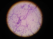
Bacteria in Photos

Bacteria in Photos