CELL DISRUPTION
Introduction
Biological products synthesized by
fermentation or cell culture are either intracellular or extracellular.
Intracellular products either occur in a soluble form in the cytoplasm or are
produced as inclusion bodies (fine particles deposited within the cells).
Examples of intracellular products include recombinant insulin and recombinant
growth factors. A large number of recombinant products are found as inclusion
bodies in order to accumulate in larger quantities within the cells. In order
to obtain intracellular products the cells first have to be disrupted to
release these into a liquid medium before further separation can be carried
out. Certain biological products have to be extracted from tissues, an example
being porcine insulin which is obtained from pig pancreas. In order to obtain
such a tissue derived substance, the source tissue first needs to be
homogenized or ground into a cellular suspension and the cells are then
subjected to cell disruption to release the product into a solution. In the
manufacturing process for intracellular products, the cells are usually first
separated from the culture liquid medium. This is done in order to reduce the
amount of impurity: particularly secreted extracellular substances and
unutilized media components. In many cases the cell suspensions are thickened
or concentrated by microfiltration or centrifugation in order to reduce the
process volume.
Several
factors must be considered.
Volume or sample size of cells to be disrupted
If
only a few microliters of sample are available, care must be taken to minimize
loss and to avoid cross-contamination.
Disruption
of cells, when hundreds or even thousands of liters of material are being
processed in a production environment, presents a different challenge.
Throughput, efficiency, and reproducibility are key factors.
How many different samples need to be disrupted at one
time?
Frequently
when sample sizes are small, there are many samples. As sample sizes increase,
fewer samples are usually processed. Issues are sample cross contamination,
speed of processing, and equipment cleaning.
How easily are the cells disrupted?
As
the difficulty of disruption increases (e.g.
E. coli),
more force is required to efficiently disrupt the cells. For even more
difficult samples (e.g.
yeast), there is a parallel increase in the processor power
and cost. The most difficult samples (e.g.
spores) require
mechanical forces combined with chemical or enzymatic efforts, often with
limited disruption efficiency.
What efficiency of disruption is required?
Over-disruption
may impact the desired product. For example, if
subcellular fractionation studies are
undertaken, it is often more important to have intact subcellular components,
while sacrificing disruption efficiency.
For
production scale processes, the time to disrupt the cells and the
reproducibility of the method become more important factors.
How stable is the molecule(s) or component that needs
to be isolated?
In
general, the cell disruption method is closely matched with the material that
is desired from the cell studies. It is usually necessary to establish the
minimum force of the disruption method that will yield the best product.
Additionally, once the cells are disrupted, it is often essential to protect
the desired product from normal biological processes (e.g. proteases) and from
oxidation or other chemical events.
What purification methods will be used following cell
disruption?
It
is rare that a cell disruption process produces a directly usable material; in
almost all cases, subsequent purification events are necessary. Thus, when the
cells are disrupted, it is important to consider what components are present in
the disruption media so that efficient purification is not impeded.
Is the sample being subjected to the method
biohazardous?
Preparation
of cell-free extracts of pathogens presents unique difficulties. Mechanical
disruption techniques are not always applicable owing to potential biohazard
problems associated with contamination of equipment and generation of aerosols.
Cells
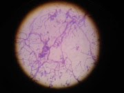
Different types of cell need to be disrupted in the bioindustry:
·
Gram positive
bacterial cells
·
Gram negative
bacterial cells
·
Yeast cell
·
Mould cells
·
Cultured
mammalian cells
·
Cultured plant
cells
·
Ground tissue
Bacterial cells:
The cell wall of Gram positive bacteria is thick and mainly composed of thick
layer of peptidoglycan layer. While the plasma or cell membrane which is made
up of phospholipids and proteins is relatively fragile. In certain cases
polysaccharide capsules may be present outside the cell wall. The cell wall of
gram positive bacteria is particularly susceptible to lysis by the antibacterial enzyme, lysozyme. Unlike gram positive bacteria,
gram negative bacteria do not have distinct cell walls but instead have multilayered
envelops. The peptidoglycan layer is
significantly thinner than in gram positive bacteria. An external layer composed of lipopolysaccharides and
proteins is usually present. Another difference with gram positive bacteria is
the presence of the periplasm layers which are two liquid filled gaps, one
between the plasma membrane and the peptidoglycan layer and the other between
the peptidoglycan layer and the external lipopolysaccharides. Periplasmic
layers also exits in gram positive bacteria but these are significantly thinner
than those in gram negative bacteria. The periplasm is important in
bioprocessing since a large number of proteins, particularly recombinant
proteins are secreted into it. An elegant way to recover the periplasmic
proteins is by the use of osmotic shock. This technique is discussed below.
Yeast/Mould cells: Yeasts which are unicellular have thick cell
walls, typically 0.1 to 0.2 microns in thickness. These are mainly composed of
polysaccharides such as glucans, mannans and chitins. The plasma membrane in a
yeast cell is composed of phospholipids and lipoproteins. Mould cells are
largely similar to yeast cells in terms of cell wall and plasma membrane
composition but are multicellular and filamentous.
Mammalian cells: Mammalian cells do not possess the cell wall and
are hence quite fragile i.e. easy to disrupt.
Plant cells: Plant cells on the other hand have very thick cell
walls mainly composed of cellulose and other polysaccharides. Cell wall
wherever present is the main barrier which needs to be disrupted to recover
intracellular products. A range of mechanical methods can be used to disrupt
the cell wall. Chemical methods when used for cell disruption are based on
specific targeting of key cell wall components. For instance, lysozyme is used
to disrupt the cell wall of gram positive bacteria since it degrades
peptidoglycan which is a key cell wall constituent. In gram negative bacteria,
the peptidoglycan layer is less susceptible to lysis by lysozyme since it is
shielded by a layer composed of lipopolysaccharides and proteins.
Cell membranes: Cell membranes or plasma membranes are composed of
phospholipids arranged in the form of a bilayer with the hydrophilic groups of
the phospholipids molecules facing outside (Figure below). The hydrophobic residues
remain inside the cell membrane where they are shielded from the aqueous
environment present both within and outside the cell. The plasma membrane can
be easily destabilized by detergents, acid, alkali and organic solvents. The
plasma membrane is also quite fragile when compared to the cell wall and can
easily be disrupted using osmotic shock i.e. by suddenly changing the osmotic
pressure across the membrane. This can be achieved simply by transferring the
cell from isotonic medium to distilled water.
Figure 1:
Plasma membrane
Cell disruption
methods can be classified into two categories: Mechanical methods and Non-mechanical
methods.
A. Mechanical Methods of Cell Disruption: uses mechanical forces to disintegrate the
cells like soli shearing, liquid shearing, pressure etc. Some methods are:
1.
Disruption in bead mill
2.
Disruption using
a rotor stator mill
3.
Homogenization
4.
Disruption using
French press
5.
Disruption using
ultrasonic vibrations
B. Non-mechanical
Methods of Cell Disruption: includes physical, chemical and biological
treatment of cells to disrupt the cells.
1. Physical methods
a. Disruption using osmotic shock
b. Freeze-thaw method
2. Chemical methods
a.
Detergents
b. Solvents
c. Acid
and alkali methods
3. Biological methods
a.
Enzymes e.g.
lysozyme
b.
Phage
c.
Autolysis
The mechanical methods are targeted more towards cell
wall disruption while the non-mechanical methods are mainly used for
destabilizing the cell membrane.
A.
Mechanical methods for cell disruption
1. Cell disruption using bead mill
Bead mill equipment consists of a tubular
vessel made of metal or thick glass within which the cell suspension is placed
along with small metal or glass beads. The tubular vessel is then rotated about
its axis and as a result of this the beads start rolling away from the
direction of the vessel rotation. At higher rotation speeds, some the beads
move up along with the curved wall of the vessel and then cascade back on the
mass of beads and cells below. The cell disruption takes place due to the
grinding action of the rolling beads as well as the impact resulting from the
cascading beads.
Figure 2:
Cell disruption by Bead Milling
Bead milling can generate enormous
amounts of heat. While processing thermolabile material, the milling can be
carried out at low temperatures, i.e. by adding a little liquid nitrogen into
the vessel. This is referred to as cryogenic bead milling. An alternative
approach is to use glycol cooled equipment. A bead mill can be operated in a
batch mode or in a continuous mode and is commonly used for disrupting yeast
cells and for grinding animal tissue. Using a small scale unit operated in a
continuous mode, a few kilograms of yeast cells can be disrupted per hour.
Larger unit can handle hundreds of kilograms of cells per hour.
Disadvantages:
- Occasional
problems with foaming and sample heating, especially for larger samples.
- Tough
tissue samples such as skin or seeds are difficult to disrupt unless the
sample is very small or has been pre-chopped into small pieces.
2. Cell disruption using rotor-stator mill
A rotor-stator mill device consists of a
stationary block with a tapered cavity called the stator and a truncated cone
shaped rotating object called the rotor. Typical rotation speeds are in the
10,000 to 50,000 rpm range. The cell suspension is fed into the tiny gap
between the rotating rotor and the fixed stator. The feed is drawn in due to
the rotation and expelled through the outlet due to centrifugal action. The
high rate of shear generated in the space between the rotor and the stator as
well as the turbulence thus generated are responsible for cell disruption.
These mills are more commonly used for disruption of plant and animal tissues
based material and are operated in the multipass mode, i.e. the disrupted
material is sent back into the device for more complete disruption. The cell
disruption caused within the rotor-stator mill can be described using the
equations discussed for a bead mill.
Figure 3:
Cell disruption using rotor-stator mill
3. Homogenization
Liquid
based homogenization is the most widely used cell disruption technique for
small volumes and cultured cells. Cells are lysed by forcing the cell or tissue
suspension through a narrow space known as clearance space (0.001 mm- 0.006 mm),
thereby shearing the cell membranes. Three different types of homogenizers are
in common use. A Dounce homogenizer consists of a round glass pestle that is
manually driven into a glass tube. A Potter-Elvehjem homogenizer consists of a
manually or mechanically driven Teflon pestle shaped to fit a rounded or
conical vessel. The number of strokes and the speed at which the strokes are
administered influences the effectiveness of Dounce and Potter-Elvehjem
homogenization methods. Both homogenizers can be obtained in a variety of sizes
to accommodate a range of volumes.
Figure 4: Cell
disruption using homogenizer
4. Cell disruption using French press
A French
press is a device commonly used for small scale recovery of intracellular
proteins and DNA from bacterial and plant cells. The device consists of a
cylinder fitted with a plunger which is connected to a hydraulic press. The
cell suspension is placed within the cylinder and pressurized using the
plunger. The cylinder is provided with an orifice through which the suspension
emerges at very high velocity in the form of a fine jet. The cell disruption
takes place primarily due to the high shear rates and differential pressure. The internal FRENCH Pressure Cell pressure
increases as the pressure developed by the Laboratory Press increases. The
intracellular pressure increases as well. As the sample is dispensed through
the sample outlet tube, the external pressure on the cell wall drops rapidly
toward atmospheric pressure. The pressure within the cell drops as well but not
as quickly as the pressure external to the cell. This pressure differential
causes the cell wall membrane to burst, releasing the intra-cellular contents. A French press is frequently provided with an impact
plate, where the jet impinges causing further cell disruption. Typical volumes
handled by such devices range from a few milliliters to a few hundred milliliters.
Typical operating pressure ranges from 10,000 to 50,000 psi.
Advantages:
— This technique results in more uniform and
complete disruption
— Cells do not require pre-treating.
— Easy to use
Figure 5:
Cell disruption using French press
5. Cell disruption using ultrasonic vibrations
Ultrasonic vibrations (i.e. having
frequency greater than 18 kHz) can be used to disrupt cells. The cells are
subjected to ultrasonic vibrations by introducing an ultrasonic vibration
emitting tip into the cell suspension (Figure below). Ultrasound emitting tips
of various sizes are available and these are selected based on the volume of
sample being processed. The ultrasonic vibration could be emitted continuously
or in the form of short pulses. A frequency of 25 kHz is commonly used for cell
disruption. The duration of ultrasound needed depends on the cell type, the
sample size and the cell concentration. These high frequency vibrations cause
cavitations, i.e. the formation of tiny bubbles within the liquid medium. When
these bubbles reach resonance size, they collapse releasing mechanical energy
in the form of shock waves equivalent to several thousand atmospheres of
pressure. The shock waves disrupt cells present in suspension. For bacterial
cells such as E. coli, 30 to 60
seconds may be sufficient for small samples. For yeast cells, this duration
could be anything from 2 to 10 minutes.
Ultrasonic vibration is frequently used
in conjunction with chemical cell disruption methods. In such cases the
barriers around the cells are first weakened by exposing them to small amounts
of enzymes or detergents. Using this approach, the amount of energy needed for
cell disruption is significantly reduced.
Disadvantages:
- Heat
generated by the ultrasound process must be dissipated.
- High noise
levels (most systems require hearing protection and sonic enclosures)
- Yield
variability
- Free
radicals are generated that can react with other molecules.
Figure 6: Cell disruption by sonication
6. Mortar and Pestle
It is
a manual grinding method, most commonly used to disrupt plant cells. In this
method, tissue is frozen in liquid nitrogen and then crushed using a mortar and
pestle. Because of tensile strength of the cellulose and other polysaccharides
comprising the cell wall, this method is the fastest and most efficient way to
access plant protein and DNA.