Table-1: Colonial morphology of common molds (Fungi)
S. no. | Fungi | Colony morphology |
1. | Penicillium | Bluish-green; brush arrangement of phialospores. |
2. | Aspergillus | Bluish-green with sulfur-yellow areas on the surface. Aspergillus |
3. | Scopulariopsis | Light brown; rough-walled microconidia. |
4. | Trichoderma | Green, resemble Penicillium macroscopically. |
5. | Gliocadium | Dark green; conidia (phialospores) borne on phialides, similar to Penicillium; grows faster than Penicillium. |
6. | Cladosporium (Hormodendrum) | Light green to grayish surface; gray to black back surface; blastoconidia. |
7. | Pleospora | Tan to green surface with brown to black back; ascospores shown are produced in sacs borne within brown, flask-shaped fruiting bodies called pseudothecia. |
8. | Scopulariopsis | Light brown; rough-walled microconidia. |
9. | Paecilomyces | Yellowish-brown; elliptical microconidia. |
10. | Alternaria | Dark greenish-black surface with gray periphery; black on reverse side; chains of macroconidia. |
11. | Bipolaris | Black surface with grayish periphery; macroconidia shown. |
12. | Pullularia | Black, shiny, leathery surface; thick walled; budding spores. |
13. | Diplosporium | Buff-colored wooly surface; reverse side has red center surrounded by brown. |
14. | Oospora (Geotrichum) | Buff-colored surface;hyphae break up into thin-walled rectangular arthrospores. |
15. | Fusarium | Variants of yellow, orange, red, and purple colonies; sickle-shaped macroconidia. |
16. | Trichothecium | White to pink surface; two-celled conidia. |
17. | Mucor | A zygomycete; sporangia with a slimy texture; spores with dark pigment. |
18 | Rhizopus | A zygomycete; spores with dark pigment. |
19. | Syncephalastrum | A zygomycete; sporangiophores bear rod-shaped sporangioles, each containing a row of spherical spores. |
20. | Nigrospora | Conidia black, globose, one-celled, borne on a flattened, colorless vesicle at the end of a conidiophore. |
21. | Montospora | Dark gray center with light gray periphery; yellow-brown conidia. |
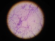
Figure-1: Microscopic appearance of some of the more common molds

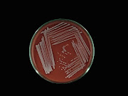
Figure-2: Colony characteristics of some of the more common molds

Source:
Benson. 2001 Microbiological Applications Laboratory Manual in General Microbiology, Eighth Edition, the McGraw-Hill Companies.


