Wednesday, September 1, 2010

Survivability of Bacillus thuringiensis in waste water

(Report to be submitted at UGC)

ABSTRACT

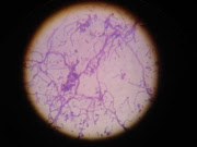
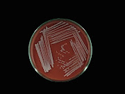
Bacillus thuringiensis strains were isolated from soil samples collected from Sagarmatha National Part of the Everest region and subsequently identified by standard microbiological techniques including colonial characteristics, morphological characteristics and biochemical characteristics. Some of potent strains already preserved in lab were also revived and recharacterized for their crystal protein production property. The stationary phase culture broth was tested for insect bioassay. All these isolates were able to kill the mosquito larva even at their natural breeding place, waste water and were further used for survivability test in the waste water. The growth of B. thuringiensis in waste water was found to be soared for seventh days and plunged gradually in subsequent days till 30 days. The survivability of B. thuringiensis isolates was recorded till 30 days and found to be alive in the waste water. The number of B. thuringiensis was found to be higher in control in compare with the test, unautoclaved waste water.

Friday, August 13, 2010

How to identify Fungi ???

Table-1: Colonial morphology of common molds (Fungi)

S. no.

Fungi

Colony morphology

1.

Penicillium

Bluish-green; brush arrangement of phialospores.

2.

Aspergillus

Bluish-green with sulfur-yellow areas on the surface. Aspergillus niger is black.

3.

Scopulariopsis

Light brown; rough-walled microconidia.

4.

Trichoderma

Green, resemble Penicillium macroscopically.

5.

Gliocadium

Dark green; conidia (phialospores) borne on phialides,

similar to Penicillium; grows faster than Penicillium.

6.

Cladosporium

(Hormodendrum)

Light green to grayish surface; gray to black back surface;

blastoconidia.

7.

Pleospora

Tan to green surface with brown to black back; ascospores shown are produced in sacs borne within brown, flask-shaped fruiting bodies called pseudothecia.

8.

Scopulariopsis

Light brown; rough-walled microconidia.

9.

Paecilomyces

Yellowish-brown; elliptical microconidia.

10.

Alternaria

Dark greenish-black surface with gray periphery; black on reverse side; chains of macroconidia.

11.

Bipolaris

Black surface with grayish periphery; macroconidia shown.

12.

Pullularia

Black, shiny, leathery surface; thick walled; budding spores.

13.

Diplosporium

Buff-colored wooly surface; reverse side has red center surrounded by brown.

14.

Oospora (Geotrichum)

Buff-colored surface;hyphae break up into thin-walled

rectangular arthrospores.

15.

Fusarium

Variants of yellow, orange, red, and purple colonies; sickle-shaped macroconidia.

16.

Trichothecium

White to pink surface; two-celled conidia.

17.

Mucor

A zygomycete; sporangia with a slimy texture; spores with dark pigment.

18

Rhizopus

A zygomycete; spores with dark pigment.

19.

Syncephalastrum

A zygomycete; sporangiophores bear rod-shaped sporangioles, each containing a row of spherical spores.

20.

Nigrospora

Conidia black, globose, one-celled, borne on a flattened,

colorless vesicle at the end of a conidiophore.

21.

Montospora

Dark gray center with light gray periphery; yellow-brown conidia.

Figure-1: Microscopic appearance of some of the more common molds


Figure-2: Colony characteristics of some of the more common molds


Source:

Benson. 2001 Microbiological Applications Laboratory Manual in General Microbiology, Eighth Edition, the McGraw-Hill Companies.

Sunday, August 8, 2010

A Practical Manual of Environmental Microbiology


Bacteria in Photos

Bacteria in Photos