Proposal on
Characterization of broad spectrum antibiotics from Actinomycetes isolates from Khumbu region for the search of novel ones.
Introduction
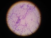
Actinomycetes comprise an extensive and diverse group of Gram-positive, aerobic, mycelial bacteria with high G+C nucleotide content (>55%), and play an important ecological role in soil cycle. The name of the group actinomycetes is derived from the first described anaerobic species Actinomyces bovis that causes actinomycosis, the ‘ray-fungus disease’ of cattle. They were originally considered to be intermediate group between bacteria and fungi but are now recognized as prokaryotic microorganisms (Kuster 1968).
The majority of Actinomycetes are free living, saprophytic bacteria found widely distributed in soil, water and colonizing plants. Actinomycetes population has been identified as one of the major group of soil population (Kuster 1968), which may vary with the soil type. They belong to the order Actinomycetales (Superkingdom: Bacteria, Phylum: Firmicutes, Class: Actinobacteria, Subclass: Actinobacteridae). According to Bergey's Manual Actinomycetes are divided into eight diverse families: Actinomycetaceae, Mycobacteriaceae, Actinoplanaceae, Frankiaceae, Dermatophilaceae, Nocardiaceae, Streptomycetaceae, Micromonosporaceae (Holt, 1989) and they comprise 63 genera (Nisbet and Fox, 1991). Based on 16s rRNA classification system they have recently been grouped in ten suborders: Actinomycineae, Corynebacterineae, Frankineae, Glycomycineae, Micrococineae, Micromonosporineae, Propionibacterineae, Pseudonocardineae, Streptomycineae and a large member of Streptomyces are still remained to be grouped (www.ncbi.nlm.nih.gov). Actinomycetes have characteristic biological aspects such as mycelial forms of growth that accumulates in sporulation and the ability to form a wide variety of secondary metabolites including most of the antibiotics.
One of the major groups in actinomycetes is Streptomyces. Streptomyces contains 69-78 mol% of G+C. Substrate and aerial mycelium is highly branched. Substrate hyphae are 0.5-1.0 µm in diameter. In the colony ages aerial mycelia develop into chain of spores (conidia) by the formation of crosswalls in the multinucleated aerial filaments. Conidial wall are convoluted projection which together with the shape and the arrangement of the spore-bearing structure are characteristic of each species of Streptomyces (Anderson et al., 2001). It produces several antibiotics including of aminoglycosides, anthracyclins, glycopeptides, b-lactams, macrolides, nucleosides, peptides, polyenes, polyethers and tetracyclines (Sahin and Ugur, 2003).
Thus investigators turn towards Streptomyces and also other genera of actinomycetes such as Nocardia, Micromonospora, Thermoactinomycetes etc. for isolation of novel antibiotics. No doubt soil is the natural habitat of most of the microorganisms where vast array of bacteria, actinomycetes, fungi and other organisms exist and provided with suitable growth condition and ability to proliferate. Thus most actinomycetes contributing to antibiotic production are screened from soil (Williams and Khan, 1974).
Our prime focus is to find out the novel antibiotic with broad-spectrum antimicrobial activity from Actinomyecetes isolates of high altitude.
Background
In, RLABB, The first work on the diversity of actinomycestes was started by Singh, D. and Agrawal, V.P. (2002). The research on actinomycetes form
Objectives
1. To subculture actinomycetes isolates which are already present in RLABB
2. To screen for antibiotic production
3. To partially purify the antibiotics
4. To characterize the antibiotics by GC analysis
Methodology
1. Isolation and Purification of Actinomycetes
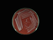
Soil samples will be obtained from Research Laboratory for Agricultural Biotechnology and Biochemistry (RLABB). Isolation of actinomycetes will be performed by soil dilution plate technique using Starch-Casein Agar (Singh and Agrawal, 2002 & 2003). Actinomycetes on the plates will be identified as colored, dried, rough, with irregular/regular margin; generally convex colony as described by Williams and Cross (1971). Streak plate method will be used to purify cultures of actinomycetes (Williams and Cross, 1971, Singh and Agrawal 2002; Agrawal 2003). After isolation of the pure colonies based on their colonial morphology, colour of hyphae, color of aerial mycelium, they will be individually plated on another but the same agar medium.
2. Morphological and Biochemical characterization
Morphological examination of the actinomycetes will be done by using cellophane tape and cover slip-buried methods (Williams and Cross, 1971; Singh and Agrawal 2002; Singh and Agrawal 2003). The mycelium structure, color and arrangement of conidiophores and arthrospore on the mycelium will be examined under oil immersion (1000X). The observed structure will be compared with Bergay’s manual of Determinative Bacteriology, Ninth edition (2000) for identification Streptomyces spp. Different biochemical tests will be performed to characterize the Streptomyces spp. The tests generally used are gelatin hydrolysis, starch hydrolysis, urea- hydrolysis, acid production from different sugars utilization tests, resistance to NaCl, temperature tolerance test, hydrogen sulphide production test, motility test, triple sugar iron (TSI) agar test, citrate utilization test, indole test, methyl red test, voges-proskauer (Acetoin Production) test, catalase test, oxidase test (Holt 1989; Singh and Agrawal 2002; Singh and Agrawal 2003).
3. Screening of Actinomycetes for antimicrobial activity
3.1 Primary screening:
Primary screening of pure isolates will be determined by perpendicular streak method on Muller Hinton agar (MHA). In vitro screening of isolates for antagonism: MHA on Nutrient Agar (NA) plates will be prepared and inoculated with Actinomycetes isolate by a single streak of inoculum in the center of the petridish. After 4 days of incubation at 28 °C the plates were seeded with test organisms (Bacillus subtilis, Staphylococcus aureus, Enterobacter aerogens, Escherichia coli, Klebsiella species, Proteus species, Pseudomonas species, Salmonella typhi and Shigella species) by a single streak at a 90° angle to Actinomycetes strains. The microbial interactions were analyzed by the determination of the size of the inhibition zone.
3.2 Secondary screening:
Secondary screening is performed by agar well method against the standard test organism. Fresh and pure culture of each strain from the primary screening will be inoculated in starch casein broth and incubated at accordingly for 7 days in water bath shaker. The visible pellets, clumps or aggregates and turbidity in the broth, will confirm growth of the organism in the flask. Contents of flasks will be filtered through Whatman no.1 filter paper. The filtrate will be used for the determination of antimicrobial activity against the standard test organisms by agar well method.
4. Antibiotics Fermentation process
Isolates showing the broad-spectrum antimicrobial activity are grown in submerged culture in 250 ml flasks containing 50 ml of broth describe in Sahin & Ugur, 2003. The flasks are inoculated with 1ml of active Actinomycetes culture and incubated at 28ºc for 7 days with shaking at 500 rpm. After fermentation, fermented broth will filtered through Whatman no.1 filter paper.
5. Extraction of antimicrobial metabolites
Antibacterial compound will be recovered from the filtrate by treating twice with one volume of ethyl acetate (Busti et al., 2006). And after evaporation residue will be used for determination of antimicrobial activity, minimum inhibitory concentration and to perform bioassay of antibiotic (Pandey et al., 2004).
6. Thin Layer Chromatography and Bioassay of antibiotic
Silica gel plates, 10 X 20 cm, 1mm thick, are prepared. They are activated at 150°C for half an hour. Ten micro-liters of the ethyl acetate fractions and reference antibiotics are applied on the plates and the chromatogram is developed using chloroform: methanol (4:1) as solvent system. The plates are run in duplicate; one set is used as the reference chromatogram and the other is used for Bioassay of antibiotic. The spots in the chromatogram are visualized in the iodine vapor chamber and UV chamber (Thangadural et al., 2002 and Pandey et al., 2004).
Results of previous research
Table 1: Total number of antibiotic producing actinomycetes
S.No. | Soil Sample from | Height (m) | Total Actinomycetes isolates | Number of antibiotic producing isolates |
1 | Lukla | 2660 | 14 | 9 |
2 | Lobuche | 5000-5300 | 12 | 7 |
3 | 3446 | 14 | - | |
4 | Jorsale | 2837 | 36 | - |
5 | Tengboche | 3867 | 8 | - |
6 | Kalapattar | >5500 | 79 | 7 antibiotic producing actinomycetes screening out of 9 isolates. |
7 | Manang | 3200-3600 | 45 | 45 |
Total | 208 | 68 |
Out of these total antibiotic producing Actinomycetes, the following are the most potent strains producing broad spectrum antibiotics (inhibiting both Gram positive bacteria including Staphycoccus aureus, Bacillus subtilis and Gram negative bacteria including E.coli, Salmonella typhii, Salmonella paratyphii, Proteus vulgaris, Proteus mirabilis, Klebseilla pneumoniae, K. oxytoca, Shigella spp). But none of isolates were found to inhibit Pseudomanas spp used as test organism in our research.
Table 2: Total number of broad spectrum antibiotic producing Actinomycetes
Isolates from Kalapathar | |
1 | K.6.3 |
2 | K.14.2 |
3 | K.58.5 |
4 | K.8.2 |
5 | K.16.4 |
6 | K.60.4 |
Isolates from Manang | |
7 | M30d |
Isolates from Lobuche | |
8. | Lob18.2b |
Expected Outcomes
Being majority of antibiotics producing bacteria are Actinomycetes (mainly Streptomyces spp.) our research work we will select different actinomycetes producing broad-spectrum antibiotics. The antibiotics will be partially purified and again bioassayed against the test organisms. The active fraction will be then analyzed for chemical characterization. Since these species are from very cold Everest region, the organisms as well as antibiotics produced by them may be novel ones.
References
Busti E, Monciardini P, Cavaletti L, Bamonte R, Lazzarini A and Sosio et al. (2006) Antibiotic-producing ability by representatives of a newly discovered lineage of actinomycetes. Microbiology 152: 675-683
Holt JG 1989 Bergey's manual of systematic bacteriology, vol 4, ed. S.T. Williams and M.E. Sharpe,
Kuster HJ, (1968) Uber die Bildung Von Huminstoffen durch Streptomyceten. Landwirtsch. Forsch
Nisbet LJ and Fox FM (1991) The importance of microbial biodiversity to biotechnology, In, The biodiversity of microorganisms and invertebrates: its role in sustainable Agriculture, ed.D.L. Hawksworth, 224-229, CAB International.
Pandey B, Ghimire P and Agrawal VP (2004) Studies on Antibacterial Activity of Soil from Khumbu Region of
Sahin N and Ugur A (2003) Investigation of the Antimicribial Activity of some Streptomyces isolates. Turk J Biol 27: 79-84.
Singh D and Agrawal VP (2002) Microbial Biodiversity of Mount Everest Region, a paper presented in International Seminar on Mountains -
Singh D and Agrawal VP (2003) Diversity of Actinomycetes of Lobuche in Mount Everest I Proceedings of International Seminar on Mountains –
Thangadural S,


0 comments:
Post a Comment